Cirujano de prótesis de cadera y rodilla en Nueva York
El Dr. Nakul Karkare es un especialista en cadera y rodilla de Nueva York. Ha realizado con éxito decenas de reemplazo de cadera & cirugías de reemplazo de rodilla en los hospitales de Manhattan a Long Island en los últimos 10+ años.
Historias de pacientes ortopédicos:
Tengo 72 años y me han reemplazado la cadera derecha. Tuve problemas durante más de un año y seguí posponiéndolo pensando que desaparecería, pero fue empeorando progresivamente.
Prótesis articular mínimamente invasiva
La cirugía mínimamente invasiva de sustitución articular utiliza pequeñas incisiones e instrumentos especialmente diseñados para realizar intervenciones sencillas y complejas sin necesidad de grandes incisiones. Las incisiones más pequeñas permiten al cirujano preservar el tejido sano para que los pacientes disfruten de menos complicaciones y de una recuperación más rápida y cómoda. El Dr. Karkare tiene una gran experiencia y habilidad en procedimientos quirúrgicos mínimamente invasivos para la sustitución articular, utilizando enfoques innovadores y la tecnología más avanzada para la colocación precisa de los componentes del implante a través de incisiones más pequeñas que preservan el tejido. Al igual que con otras técnicas quirúrgicas y enfoques, la decisión de utilizar un enfoque quirúrgico mínimamente invasivo se toma sobre una base de paciente por paciente para ayudar a asegurar que cada paciente logra los mejores resultados posibles a largo plazo.
Prótesis robótica de cadera
La sustitución articular robótica puede parecer sacada de una novela de ciencia ficción, pero gracias a los recientes avances tecnológicos, la asistencia robótica se ha convertido en un método quirúrgico muy eficaz utilizado para mejorar la precisión quirúrgica y proporcionar a los pacientes recuperaciones más rápidas y mejores resultados a largo plazo. En la cirugía articular asistida por robot, se utiliza tecnología informática para ayudar al cirujano a controlar mejor aspectos muy precisos del proceso de sustitución articular, lo que permite una colocación más exacta de los implantes y una alineación ideal de los componentes de la articulación artificial. En sus consultas, el Dr. Karkare confía en el avanzado sistema asistido por robot MAKOplasty® para lograr una mejor colocación de los implantes y resultados reproducibles en función de la anatomía única de cada paciente. Las cirugías de prótesis de cadera asistidas por robot se realizan mediante un abordaje quirúrgico anterior, un método que no daña los tejidos y permite una recuperación más rápida y cómoda.
Prótesis anterior de cadera
Tradicional prótesis de cadera utiliza grandes incisiones a través de los principales grupos musculares de la zona posterior de la cadera para acceder a la articulación de la cadera dañada, lo que provoca un mayor daño tisular y un periodo de recuperación más largo y menos cómodo. En la artroplastia anterior de cadera, las incisiones se realizan a lo largo de la parte delantera (o anterior) de la cadera. Estas incisiones pueden situarse con precisión para evitar daños importantes en los tejidos, apartando los músculos en lugar de cortarlos. Los pacientes que son buenos candidatos para una artroplastia de cadera por vía anterior pueden beneficiarse de una cicatrización y recuperación más rápidas, menos molestias postoperatorias y un retorno más rápido a las actividades normales y a una función óptima de la articulación, en comparación con los hombres y mujeres que se someten a artroplastias de cadera tradicionales.
Sustitución articular asistida por ordenador
Las técnicas quirúrgicas asistidas por ordenador se desarrollaron para permitir a los cirujanos alcanzar altos niveles de precisión en una amplia gama de procedimientos quirúrgicos, incluidas las cirugías de prótesis articulares, en las que la precisión es la clave del éxito a largo plazo y de una función y un rendimiento normales y cómodos de la articulación. Los ordenadores proporcionan a los cirujanos información continua, incluidas imágenes bidimensionales y tridimensionales en tiempo real durante el procedimiento quirúrgico, para ayudar a guiar la colocación de los componentes del implante en función de la anatomía única del paciente y otros factores. El Dr. Karkare cuenta con una amplia experiencia en técnicas de sustitución articular asistidas por ordenador, utilizando los sistemas y métodos quirúrgicos más avanzados para lograr una colocación precisa y exacta de los componentes articulares artificiales con el fin de conseguir una función articular óptima.
Sustitución total de rodilla
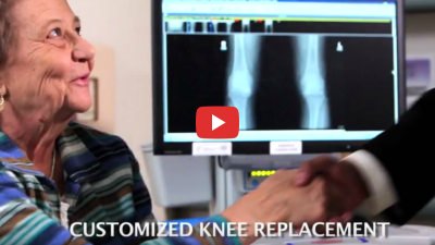
La artroplastia de rodilla ha tenido una gran demanda en los últimos años, a medida que los «baby boomers» entran en la tercera edad y siguen llevando una vida más activa. El reemplazo de la articulación de la rodilla se realiza más comúnmente en hombres y mujeres que han tenido daños en las articulaciones relacionados con la artritis, pero también se puede realizar después de una lesión significativa en la articulación de la rodilla. Dr. Karkare ofrece cirugías de reemplazo de rodilla personalizado basado en una gran cantidad de datos de diagnóstico para facilitar una mayor precisión durante la cirugía y mejores resultados después. La cirugía de prótesis de rodilla a medida ofrece a los pacientes muchas ventajas, como un ajuste mejor y más estable, una recuperación más rápida tras la intervención, menos dolor durante la recuperación y menos riesgos de complicaciones como la infección. La prótesis de rodilla a medida también daña menos los tejidos, por lo que los pacientes pueden reanudar sus actividades normales más rápidamente. Prótesis total de rodilla es la más habitual, en la que se extrae y sustituye el hueso dañado tanto del fémur como de la tibia (huesos de la parte superior e inferior de la pierna) para restablecer la amplitud de movimiento sin dolor. En pacientes con lesiones articulares menos extensas puede considerarse la sustitución parcial de la rodilla.
Cirugías de sustitución articular
La cirugía de prótesis articular puede proporcionar a hombres y mujeres una mayor movilidad y una mejora significativa de la calidad de vida, pero es una decisión que requiere una cuidadosa consideración y la orientación de un especialista en prótesis articular cualificado y con experiencia. Antes de someterse a una operación de prótesis articular, todos los pacientes se someten a una evaluación diagnóstica exhaustiva y completa para descartar enfoques terapéuticos no quirúrgicos más conservadores. La cirugía se considera cuando se ha determinado que las opciones de tratamiento más conservadoras son ineficaces o inadecuadas para aliviar el dolor y otros síntomas. Como un cirujano ortopédico de alto rango, el Dr. Karkare es experto tanto en técnicas quirúrgicas tradicionales «abiertas» y los enfoques mínimamente invasivos.
Revisión de prótesis de rodilla
Revisión de prótesis de rodilla es lo que llamamos una operación para revisar su prótesis de rodilla existente. Puede tratarse de un pequeño ajuste o de una operación masiva en la que se sustituye una gran parte del hueso. En una prótesis de rodilla típica se utiliza plástico para sustituir los extremos del hueso del muslo (fémur) y de la espinilla (tibia), insertando el plástico entre ellos y la rótula.
Necrosis avascular
También llamada osteonecrosis o necrosis aséptica, necrosis avascular es una afección grave que ocurre cuando el tejido óseo comienza a debilitarse y morir como resultado de un flujo sanguíneo inadecuado al tejido. Aunque muchas personas nunca han oído hablar de la necrosis avascular, es una de las causas más comunes de dolor de rodilla relacionado con la edad. La necrosis avascular puede ocurrir en cualquier parte, pero afecta con mayor frecuencia a las caderas y las rodillas, seguidas de los tobillos, los hombros y la parte superior de los brazos. La causa subyacente de la necrosis avascular no se comprende claramente, y los estudios de imágenes en profundidad combinados con la experiencia y los conocimientos profesionales desempeñan un papel fundamental en el diagnóstico de la afección. En última instancia, el objetivo del tratamiento es detener la progresión de la enfermedad y restaurar el uso normal y sin dolor de la articulación o extremidad. Si bien la intervención no quirúrgica puede brindar un alivio temporal de los síntomas, la mayoría de los pacientes requerirán cirugía para restaurar las áreas dañadas y aliviar el dolor.
Hip Bursitis
La bursitis de cadera ocurre cuando los pequeños sacos llenos de líquido llamados bursa se inflaman, a veces como resultado de una lesión, cirugía, deportes de movimiento repetitivo como correr, enfermedad de la columna, artritis reumatoide o anomalías anatómicas en la pierna o la articulación. La bursitis de cadera a menudo se trata sin cirugía, con medicamentos y descanso para ayudar a reducir la inflamación y aliviar el dolor. Los casos más avanzados o «obstinados» de bursitis de cadera pueden requerir cirugía para extirpar la bursa inflamada sin limitar el movimiento normal de la articulación. El Dr. Karkare evalúa a cada paciente individualmente para garantizar que se brinde la atención más adecuada, incluidas las opciones quirúrgicas y no quirúrgicas.
Hip Dysplasia
La displasia de cadera es una enfermedad dolorosa que suele aparecer en niños pequeños y adolescentes mientras la articulación de la cadera y los tejidos circundantes crecen y se desarrollan. Mientras que una cadera normal crece hasta desarrollar un ajuste perfecto entre la parte esférica de la articulación y su cavidad en forma de copa, en la displasia de cadera la articulación está malformada, lo que da lugar a una unión menos segura entre la bola y la copa que permite que la cadera se salga de la alineación o se disloque. La displasia de cadera puede tratarse de forma conservadora en algunos pacientes jóvenes, pero en otros casos, incluidos los pacientes que han tenido múltiples correcciones sin éxito, la cirugía de prótesis de cadera puede ser la mejor opción para el alivio a largo plazo de los síntomas y el funcionamiento normal de la articulación.
¿Por qué elegir al Dr. Karkare?
Como cirujano mejor calificado en Nueva York, el Dr. Karkare tiene una reputación estelar, con una amplia experiencia para garantizar que tendrá la más amplia gama de opciones de tratamiento seguras y efectivas para aliviar su dolor y otros síntomas. Dr. Karkare ofrece programación en múltiples ubicaciones en Manhattan y Long Island, Nueva York. Elija la ubicación más conveniente y programe ahora, para una consulta sin compromiso. Nos gusta que cada paciente tome una decisión informada y educada para que pueda sentirse seguro de su atención en cada paso del camino. Para programar su evaluación, llame al (516) 735-4032 o use nuestro formulario de contacto en línea para obtener más información.


