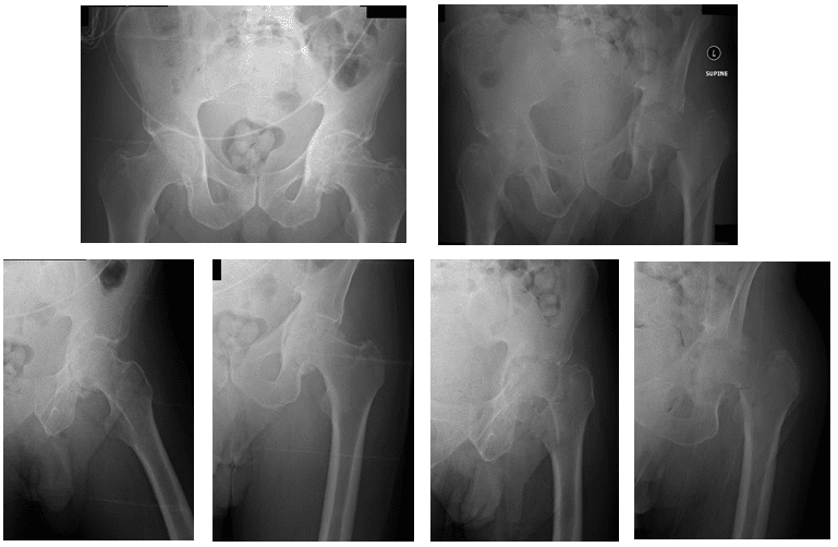
Patient is a 55 year old male who came in complaining of bilateral hip pain that he stated had been increasing over the past several months. Patient came in with x-rays to review, which indicated that he had bilateral arthritis of the hip joints. X-rays are shown below.

X-Rays show bilateral hip arthritis with worse findings of arthritis in the left hip joint
X-Rays also show severe erosive change of the left hip and a basicervical fracture at the femoral neck. Also shown is mild axial migration of the femoral head of the right hip.
To check for an infection, patient was advised to get an aspiration of the left hip. Results came back positive for infection. Patient was advised to receive bilateral Total Hip Arthroplasty (THA) with high dose antibiotic spacers to treat both the arthritis and the infection. All options and alternatives, along with the possible risks were discussed with the patient at length. Patient had decided to proceed with the surgery.

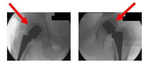
Intraoperative radiographs. Arrows show retractors

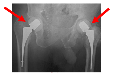
Intraoperative control films. Images show cement spacer placement of both femoral heads and necks presumably. X-Rays show high dose antibiotic spacers

X-Ray shows pelvis post bilateral hip cement placer placement. X-Rays show cement spacers

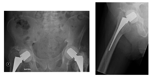
X-Rays show bilateral hip spacer with no loosening or pelvic fractures
Patient had continued to follow-up every few months to monitor his progress. He has subsequently presented pain free with good range of motion and is weight bearing as tolerated.
*Patient identifiers and dates changed to protect patient privacy.


