Patient is an 80 year old female who had come in from out of state after being advised to receive an amputation of her left leg. Patient previously had 5 surgeries, including irrigation, debridement, and drainage of her left knee after she was involved in a car accident several years ago. Previous surgeries were performed by anout of state facility.
Patient came in with X-rays to review, as shown below. Upon examination, patient was experiencing pain and was ultimately advised to receive a left staged Total Knee Arthroplasty (TKA) Reconstruction surgery. All alternatives and options were discussed at length with the patient.
It was explained to the patient that she had a comminuted fracture in which the bone broke into many pieces and that the two options were an Open Reduction Internal Fixation (ORIF) procedure or a replacement, but was advised that the replacement was the better option to achieve better results.
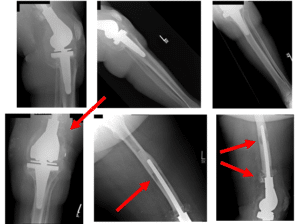
X-Rays show osteolysis of the femoral and tibial component of the left TKA prosthesis
There is also capsular calcification and capsular distention along with severe thinning of the cortex around the upper aspect of the femur. Arrows show osteolysis of the bone.

X-Rays show preoperative changes of left TKA reconstruction
Patient received an aspiration of the left knee to check for infection. Results showed that the patient had an infected knee tumor prosthesis. Patient was advised to receive an articulating antibiotic prosthesis with high dose articulating antibiotic spacers to treat the infection.
Antibiotic spacers will be left in for approximately 6-8 weeks and will then be removed and replaced with a new prosthesis. Patient had decided to proceed with the surgery.
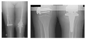
Pre-operative Scanogram show mild degenerative changes at both hips and right knee
There is an orthopedic plate and multiple screws in the right distal femur.
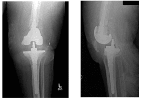
Pre-Surgical X-Rays show left TKA dislocation with dorsal translation of the tibia and widening of the tibiofemoral joint
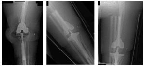
X-Rays show left knee intraoperative changes including long stem femoral and tibial prosthesis

Intraoperative Fluoroscopy Images
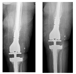
X-Rays show left knee TKA one year post-operatively
Upon follow-up patient complains of no pain, has a good range of motion, is weight-bearing as tolerated, and has been doing well subsequently.

Modular tibial tray with metaphyseal sleeve
A image above is showing a modular tibial tray used in revision knee replacement surgery. The modular tray allows attachment of a stem and metal bone augments.

Modular femoral component
The image shows a modular femoral component (posterior stabilized). The modularity of the implant allows attachment of the stem which helps transmit the load from the joint.

Metaphyseal sleeve (trabecular) with metal bone augments
The bone augments and metaphyseal sleeves help fill in the bone gap encountered during the surgery. During the extraction of the primary implant or due to infection/loosening, there may a bone gaps.
*Patient identifiers and dates changed to protect patient privacy.


